Home » Without Label » Upper Leg Tendon Anatomy / Upper Legs - Running Anatomy (Sports Anatomy) : The rectus femoris is one of the quadriceps muscles, the largest group of muscles on the front of the thigh.
Upper Leg Tendon Anatomy / Upper Legs - Running Anatomy (Sports Anatomy) : The rectus femoris is one of the quadriceps muscles, the largest group of muscles on the front of the thigh.
Upper Leg Tendon Anatomy / Upper Legs - Running Anatomy (Sports Anatomy) : The rectus femoris is one of the quadriceps muscles, the largest group of muscles on the front of the thigh.. The knee joint is commonly injured, so understanding its anatomy can help you understand the conditions that cause problems, so you stay safe and prepared. It is part of the lower limb. Notice the upper leg has a biceps muscle just like the upper arm does. This important tendon in the back of the calf and ankle stores the elastic energy needed for running, jumping, and other physical activity. Another large hip flexor is the rectus femoris.
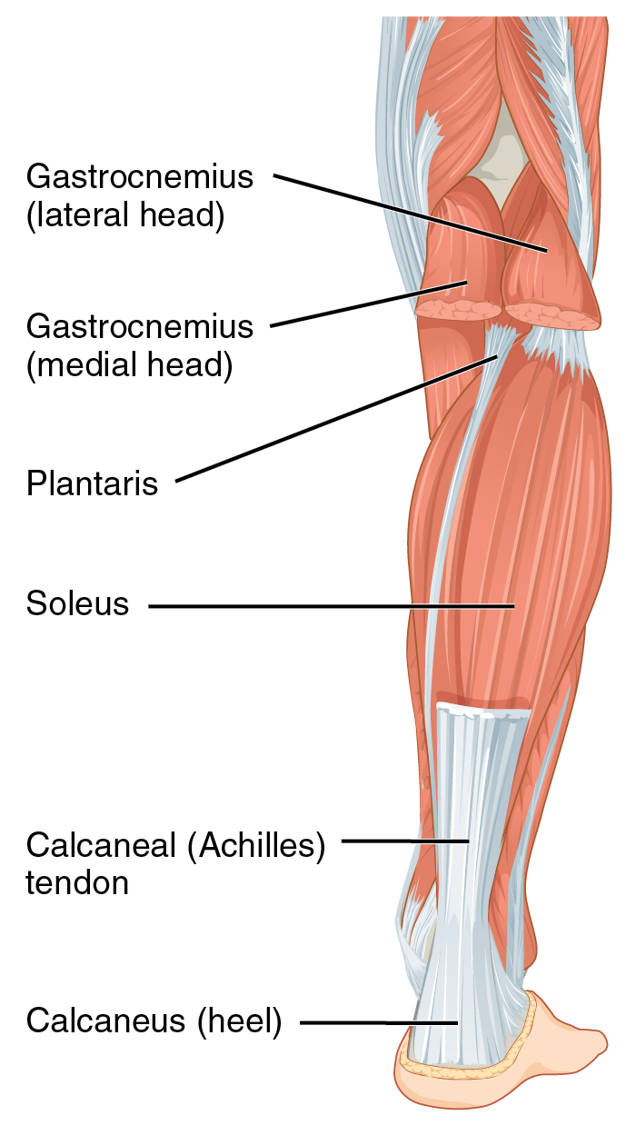
Attachment, nerve supply & action. It is a deep muscle that originates from the lower back and pelvis, and extends up to the inside surface of the upper part of the femur. The legs include the upper leg, knee, lower leg, ankle, and. The calf comprises of 2 major muscles (gastrocnemius and soleus) both of which. The hamstring muscles in the back of the thigh, the quadriceps muscles in the front, and the adductor muscles on the inside.
Achilles tendon - Wikiwand from upload.wikimedia.org Thigh muscle strain anatomy the thigh has three sets of strong muscles: The thigh has three sets of strong muscles: A muscle strain (muscle pull or tear) is a common injury, particularly among people who participate in sports. Notice the upper leg has a biceps muscle just like the upper arm does. The four muscles all extend the lower leg. The knee joint is commonly injured, so understanding its anatomy can help you understand the conditions that cause problems, so you stay safe and prepared. The hamstring muscles in the back of the thigh, the quadriceps muscles in the front, and the adductor (groin) muscles on the inside. This is why you have to indicate which biceps you are taking about when discussing one or other of these muscles.
The legs are the lower limbs of the human body that provide support and stability in addition to allowing movement.
The vastus lateralis is a muscle located on the lateral, or outside, part of your thigh. This muscle is located in front of the hip joint and provides flexion. The hamstring portion of the adductor magnus has a similar action to these muscles, but is located in the medial thigh. It runs straight down the leg and attaches to the patella via the quadriceps femoris tendon. The largest muscle masses in the leg are present in the thigh and the calf. They also are hip flexors. Possibly the most important tendon in terms of mobility is the achilles tendon. The patella is attached to the shinbone (tibia) by the patellar tendon. The hamstrings are three muscles at the back of the thigh that affect hip and knee movement. The muscles of the anterior thigh consist of the quadriceps (or quads): This deep muscle begins in the low back and pelvis and connects on the inside edge of the upper femur. The rectus femoris is located in the center of the thigh, while the vastus medialis is in the middle of the said body part. It contains many muscles and nerves but only has one bone, the femur, which is the longest and strongest bone in.
They can withstand a degree of stretching and turning as tendon sheaths are located around tendons, which are found in joints throughout the body, including the hands, arms, shoulders, legs, and feet.the human leg, in the general word sense, is the entire lower limb of the human body. Tendons are thick bands of tissue that connect muscles to bone. Tendons are cords made of tough tissue, and they work as special connector pieces between bone and muscle. The only muscle of the quadriceps to cross both the hip and knee joints. The single bone in the thigh region is called the femur.
Anatomy Of Leg Muscles And Tendons Anatomy Diagram Leg ... from i.pinimg.com This mri wrist coronal cross sectional anatomy tool is absolutely free to use. It's the area that runs from the hip to the knee in each leg. Anatomy the four quadriceps muscles meet just above the kneecap (patella) to form the quadriceps tendon. Possibly the most important tendon in terms of mobility is the achilles tendon. The rectus femoris is located in the center of the thigh, while the vastus medialis is in the middle of the said body part. This muscle is located in front of the hip joint and provides flexion. It is thin and flattened, broad above, narrow and tapering below. The hamstring portion of the adductor magnus has a similar action to these muscles, but is located in the medial thigh.
Pin on upper leg muscle anatomy from i.pinimg.com the human leg, in the general word sense, is the entire lower limb of the human body, including the foot, thigh and even the hip or gluteal region.
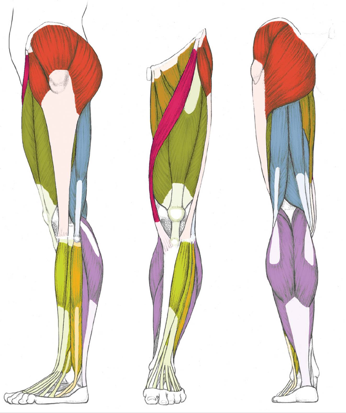
It serves to attach the plantaris, gastrocnemius (calf) and soleus muscles to the calcaneus (heel) bone. Your lower leg includes three main muscles, located behind your tibia or shinbone. The hamstring muscles in the back of the thigh, the quadriceps muscles in the front, and the adductor muscles on the inside. This muscle is located in front of the hip joint and provides flexion. For a more detailed anatomy of the muscle, check out the following leg muscle diagrams posted below. The thigh has three sets of strong muscles: The calf muscles are pivotal to movement of the ankle, foot, and toes. The hamstrings are three muscles at the back of the thigh that affect hip and knee movement. On the medial edge of the posterior thigh is the gracilis muscle. Like the biceps brachii in the arm, the biceps femoris muscle has two heads. Anatomy the four quadriceps muscles meet just above the kneecap (patella) to form the quadriceps tendon. The quadriceps tendon attaches the quadriceps muscles to the patella. The rectus femoris is located in the center of the thigh, while the vastus medialis is in the middle of the said body part.
On the medial edge of the posterior thigh is the gracilis muscle. Like the biceps brachii in the arm, the biceps femoris muscle has two heads. The calf comprises of 2 major muscles (gastrocnemius and soleus) both of which. Also called the thigh bone, this is the longest bone in the body.it. The hamstrings are three muscles at the back of the thigh that affect hip and knee movement.
Muscles of the Leg and Foot - Classic Human Anatomy in ... from doctorlib.info The legs are the lower limbs of the human body that provide support and stability in addition to allowing movement. The hamstring muscles in the back of the thigh, the quadriceps muscles in the front, and the adductor muscles on the inside. This is why you have to indicate which biceps you are taking about when discussing one or other of these muscles. Upper leg tendon anatomy : Tendons are thick bands of tissue that connect muscles to bone. Also called the thigh bone, this is the longest bone in the body.it. On the medial edge of the posterior thigh is the gracilis muscle. Anatomy the four quadriceps muscles meet just above the kneecap (patella) to form the quadriceps tendon.
The legs are the lower limbs of the human body that provide support and stability in addition to allowing movement.
The only muscle of the quadriceps to cross both the hip and knee joints. It also is active in maintaining thigh and kneecap position while walking and. Anatomy the four quadriceps muscles meet just above the kneecap (patella) to form the quadriceps tendon. Another large hip flexor is the rectus femoris. Notice the upper leg has a biceps muscle just like the upper arm does. The vastus lateralis is a muscle located on the lateral, or outside, part of your thigh. The calf comprises of 2 major muscles (gastrocnemius and soleus) both of which. The patella is attached to the shinbone (tibia) by the patellar tendon. Your upper leg includes seven major muscles. For a more detailed anatomy of the muscle, check out the following leg muscle diagrams posted below. Also called the thigh bone, this is the longest bone in the body.it. It is part of the lower limb. Smaller muscles going from the pelvis to the hip help to stabilize and rotate the hip.